Bone Cross Section Diagram - Figure 1 From Aging And The Muscle Bone Relationship Semantic Scholar : There are trabeculae in spongy bone which gives its sponge like appearance.
Bone Cross Section Diagram - Figure 1 From Aging And The Muscle Bone Relationship Semantic Scholar : There are trabeculae in spongy bone which gives its sponge like appearance.. This image added by admin. Cross section of bone diagram. So, this is the cross section of skeletal muscle (skeletal muscle of tongue). Browse 4,294 bone cross section stock photos and images available, or search for human bone cross section to find more great stock photos and pictures. A cross section of a human long bone.
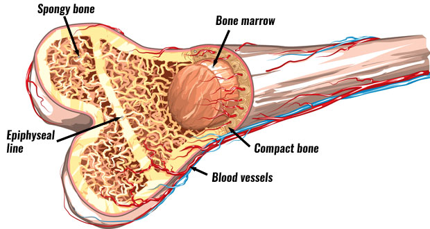
This is a cross section through decalcified bone. Bone marrow is the soft, highly vascular and flexible connective tissue within bone cavities which serve as the primary site of new blood cell production or bone marrow is the primary source of pluripotent stem cells that give rise to all hemopoietic cells (blood cells) including lymphocytes. Skeletal muscle histology description with labeled diagram well, let's me describe the skeletal muscle histology with labeled diagram so that you might understand every single structure. A cross section of a human long bone. It seems confusing and misleading.
Bone Structure Anatomy Explained What Is Bone Marrow from www.teachpe.com New users enjoy 60% off. Download 706 bone cross medical section stock illustrations, vectors & clipart for free or amazingly low rates! (b) in this micrograph of the osteon, you can see the concentric lamellae around the central canals. The star of the show (brain) is easily recognizable because it appears highly convoluted, full of ridges (gyri) and indentations (sulci).the paired thalami appear as two circular masses in the midline, forming the walls of the third ventricle.the neurocranium appears as a meshwork (trabecular. (b) in this micrograph of the osteon, you can clearly see the concentric lamellae and central canals. Cross section through the thalamus: So, this is the cross section of skeletal muscle (skeletal muscle of tongue). Browse 4,294 bone cross section stock photos and images available, or search for human bone cross section to find more great stock photos and pictures.
Anatomynote.com found full pelvis cross section horizontal female diagram from plenty of anatomical pictures on the internet.
Smartdraw includes 1000s of professional healthcare and anatomy chart templates that you can modify and make your own. (b) in this micrograph of the osteon, you can clearly see the concentric lamellae and central canals. Human bone, cross section diagram of femur showing osteon, veins, marrow. Cross section through the thalamus: This image added by admin. The bladder, like the stomach, is an expandable sac that contracts with inner folds when it is empty. Download 706 bone cross medical section stock illustrations, vectors & clipart for free or amazingly low rates! New users enjoy 60% off. You can click the image to magnify if you cannot see clearly. Related posts of cross section of a long bone bone test anatomy and physiology. The star of the show (brain) is easily recognizable because it appears highly convoluted, full of ridges (gyri) and indentations (sulci).the paired thalami appear as two circular masses in the midline, forming the walls of the third ventricle.the neurocranium appears as a meshwork (trabecular. (b) in this micrograph of the osteon, you can clearly see the concentric lamellae and central canals. Contact your company to license this image.
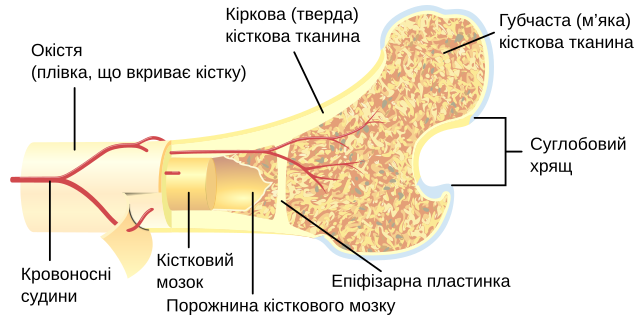
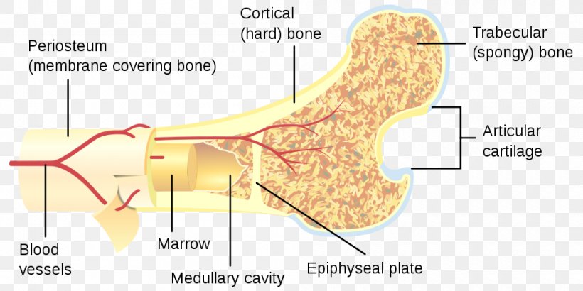
New users enjoy 60% off. The bladder, like the stomach, is an expandable sac that contracts with inner folds when it is empty. Cross section of bone diagram. Diagram orienting yourself within such a cross section is easy. Diagram with articular cartilage, marrow, medullary cavity and periosteum.
File Bone Cross Section Uk Svg Wikimedia Commons from upload.wikimedia.org (b) in this micrograph of the osteon, you can clearly see the concentric lamellae and central canals. Related posts of cross section of human bone diagram bone in arm pictures. We hope you can get the exact. Bone test anatomy and physiology 12 photos of the bone test anatomy and physiology anatomy and physiology bone lab test, anatomy and physiology bone markings test, anatomy and physiology bone practical test, anatomy and physiology bone tissue test, anatomy and physiology test on bone tissue, bone, anatomy and. Skeletal muscle histology description with labeled diagram well, let's me describe the skeletal muscle histology with labeled diagram so that you might understand every single structure. Anatomynote.com found full pelvis cross section horizontal female diagram from plenty of anatomical pictures on the internet. Thank you for visit anatomynote.com. Browse 4,294 bone cross section stock photos and images available, or search for human bone cross section to find more great stock photos and pictures.
For example, to read this diagram literally, since the cartilage can be seen inside the cutaway section of bone, it incorrectly indicates that the cartilage in fact goes through the bone structure, rather than just being found around the bone end.
Smartdraw includes 1000s of professional healthcare and anatomy chart templates that you can modify and make your own. For example, to read this diagram literally, since the cartilage can be seen inside the cutaway section of bone, it incorrectly indicates that the cartilage in fact goes through the bone structure, rather than just being found around the bone end. (b) in this micrograph of the osteon, you can clearly see the concentric lamellae and central canals. Related posts of cross section of human bone diagram bone in arm pictures. Thank you for visit anatomynote.com. Bone in arm pictures 12 photos of the bone in arm pictures bone cancer arm pictures, pictures of bone cancer in arm, bone, bone cancer arm pictures, pictures of bone cancer in arm. The inner lining of the bladder tucks into the folds and expand out to. The bladder, like the stomach, is an expandable sac that contracts with inner folds when it is empty. You can click the image to magnify if you cannot see clearly. Jump to navigation jump to search. (b) in this micrograph of the osteon, you can clearly see the concentric lamellae and central canals. Cross section of bone diagram. There are trabeculae in spongy bone which gives its sponge like appearance.
The inner lining of the bladder tucks into the folds and expand out to. The star of the show (brain) is easily recognizable because it appears highly convoluted, full of ridges (gyri) and indentations (sulci).the paired thalami appear as two circular masses in the midline, forming the walls of the third ventricle.the neurocranium appears as a meshwork (trabecular. Cross section of a long bone. This is a cross section through decalcified bone. Diagram orienting yourself within such a cross section is easy.
Long Bone Human Skeleton Fraudbein Cross Section Png 1000x500px Watercolor Cartoon Flower Frame Heart Download Free from img.favpng.com Diagram with articular cartilage, marrow, medullary cavity and periosteum. Download 706 bone cross medical section stock illustrations, vectors & clipart for free or amazingly low rates! Diagram orienting yourself within such a cross section is easy. (b) in this micrograph of the osteon, you can see the concentric lamellae around the central canals. So, this is the cross section of skeletal muscle (skeletal muscle of tongue). Shop bone cross section diagram label created by chartsanddiagrams. Cross section through the thalamus: Smartdraw includes 1000s of professional healthcare and anatomy chart templates that you can modify and make your own.
Contact your company to license this image.
The bladder, like the stomach, is an expandable sac that contracts with inner folds when it is empty. Diagram orienting yourself within such a cross section is easy. It seems confusing and misleading. Diagram with articular cartilage, marrow, medullary cavity and periosteum. The inner lining of the bladder tucks into the folds and expand out to. Jump to navigation jump to search. Skeletal muscle histology description with labeled diagram well, let's me describe the skeletal muscle histology with labeled diagram so that you might understand every single structure. Bone test anatomy and physiology 12 photos of the bone test anatomy and physiology anatomy and physiology bone lab test, anatomy and physiology bone markings test, anatomy and physiology bone practical test, anatomy and physiology bone tissue test, anatomy and physiology test on bone tissue, bone, anatomy and. Bone marrow is the soft, highly vascular and flexible connective tissue within bone cavities which serve as the primary site of new blood cell production or bone marrow is the primary source of pluripotent stem cells that give rise to all hemopoietic cells (blood cells) including lymphocytes. (b) in this micrograph of the osteon, you can clearly see the concentric lamellae and central canals. For example, to read this diagram literally, since the cartilage can be seen inside the cutaway section of bone, it incorrectly indicates that the cartilage in fact goes through the bone structure, rather than just being found around the bone end. This diagram depicts anatomy of the human eye cross section view.human anatomy diagrams show internal organs, cells, systems, conditions, symptoms and sickness information and/or tips for healthy living. New users enjoy 60% off.
This diagram depicts anatomy of the human eye cross section viewhuman anatomy diagrams show internal organs, cells, systems, conditions, symptoms and sickness information and/or tips for healthy living bone cross section. Bone marrow is the soft, highly vascular and flexible connective tissue within bone cavities which serve as the primary site of new blood cell production or bone marrow is the primary source of pluripotent stem cells that give rise to all hemopoietic cells (blood cells) including lymphocytes.


0 Komentar